KERATOCONUS TREATMENT MELBOURNE
A PATIENT’S GUIDE
Our ophthalmologists take the time to explain each diagnosis in clear, simple terms and discuss all available treatment options. We encourage questions, so you can make educated decisions about your eye care treatment with confidence.
All the specialists use cutting edge, proven technology and strive to provide you with high quality care.
HAWTHORN EYE CLINIC
What is keratoconus?
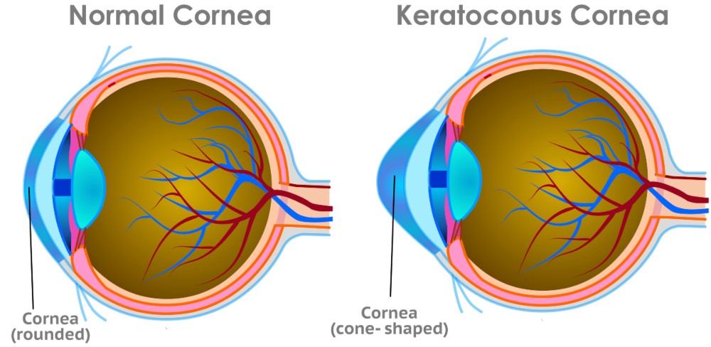
Keratoconus is a progressive condition of the cornea—the clear, dome-shaped surface at the front of the eye. Instead of maintaining its normal rounded shape, the cornea gradually becomes weaker and bulges forward in the middle into a cone-like shape. This can cause blurred, distorted or fluctuating vision and increasing sensitivity to light.
How does keratoconus affect vision?
As keratoconus develops the following affects may occur:
How is keratoconus diagnosed?
Ophthalmologists diagnose keratoconus through a comprehensive eye examination that may include:
| Diagnostic test | Description |
|---|---|
| Eye Exam: | Evaluates how clearly you see and inspects the inner eye health. |
| Corneal Topography: | Maps the cornea to help detect changes in shape & identify keratoconus. |
| Corneal Tomography: | Provides a three-dimensional corneal image to measure the front, back & thickness. |
| Pachymetry: | Measures corneal thickness, helps determine condition severity & treatments. |
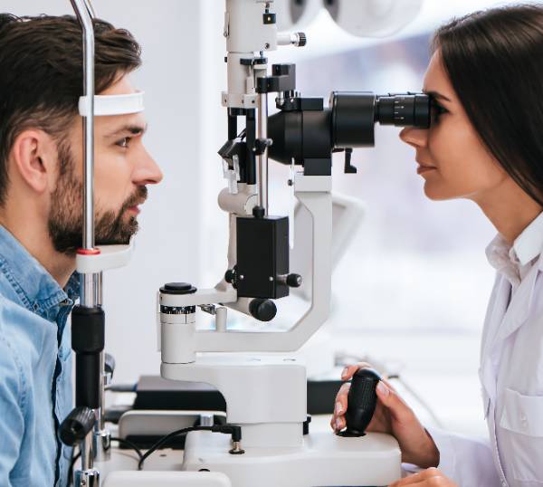
| Slit Lamp Examination | Helps examine the cornea under high magnification. |
Together, these tests help the ophthalmologist determine if keratoconus is present. Keratoconus is often progressive, repeat scans may be performed over several months to determine whether the condition is worsening.
Detecting progression is particularly important, as potential treatments may be more effective before significant corneal damage has occurred.
How is keratoconus treated?
Non-surgical options
Early keratoconus may be managed with prescription glasses or specialised contact lenses
Corneal Allogenic Intrastromal Ring Segments (CAIRS)
Corneal Allogenic Intrastromal Ring Segments (CAIRS) is a treatment for selected patients with keratoconus that uses small segments of healthy donor corneal tissue to help reshape an irregular cornea.
During the procedure, a laser is used to create precise channels within the cornea. The donor corneal segments are then placed into these channels, where they gently reshape and flatten the cone-shaped cornea, making its surface more regular. This may improve the quality of vision and may make glasses or contact lenses more effective or easier to tolerate.
Corneal Collagen Crosslinking Surgery
Corneal collagen crosslinking (CXL) is a procedure used to strengthen the cornea to potentially slow or stop the progression of keratoconus. Crosslinking strengthens the collagen fibres within the cornea, helping it become more stable and resistant to further bulging.
How does corneal crosslinking work – During the procedure, the eye is numbed with local anaesthetic drops. Riboflavin (vitamin B2) eye drops are then applied to the cornea. Riboflavin is activated by ultraviolet A (UVA) light. When the UVA light is applied, it activates the riboflavin and triggers a reaction that forms additional bonds between the collagen fibres in the cornea.
These stronger bonds increase the strength and stability of the cornea, helping it resist further weakening and bulging caused by keratoconus. Riboflavin also helps protect the deeper structures of the eye during treatment.
There are different crosslinking techniques available, and your ophthalmologist will discuss the most appropriate approach for your individual eye.
Potential Benefits of Corneal Surgery
The primary goal of corneal surgery in relation to keratoconus is to preserve vision by preventing further weakening of the cornea.
Potential benefits include:
While some patients notice mild improvements in vision, surgery does not reverse existing corneal damage and should not be considered a cure for keratoconus.
Frequently asked questions about keratoconus surgery?
Proud member of the Royal Australian and New Zealand College of Ophthalmologists.